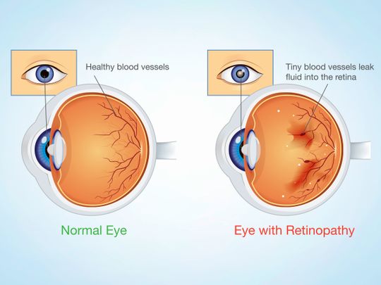
As we all know, diabetes mellitus (DM) is now a global epidemic. Diabetic retinopathy (DR) is the specific microvascular complication of DM, affecting one in three people globally.
DR remains a leading cause of vision loss in adults with DM. However, patients can reduce the risk of developing retinopathy and slow its progression by controlling blood glucose, blood pressure and blood lipids.
Risk factors
Duration of diabetes is the most critical factor in the development of DR, with 30 per cent of DM patients developing it in the first ten years of getting the disease, while many develop it in the next 30 years. Women are more prone to getting DR. Poor diabetic control, smoking, obesity and hyperlipidaemia are some of the other risk factors of the condition.

DR remains a leading cause of vision loss in adults with DM. However, patients can reduce the risk of developing retinopathy and slow its progression by controlling blood glucose, blood pressure and blood lipids.
Diagnosis
We usually ask the patient to go for a urine examination, blood sugar estimation and fundus fluorescein angiography to diagnose and monitor DR.
Ocular Coherence Tomography (OCT) — a recent advance in the diagnosis of DR — has a significant role in the diagnosis and management of DR and its complications. The technology has evolved from low resolution time domain OCT to fourier domain to the current generation of swept source OCT, which can scan the retina to a great detail.
OCT angiography is a relatively new, safe and non-invasive imaging technique, slowly replacing conventional fluorescein angiography for the diagnosis and management of DR.
Line of treatment
Treatment for DR usually begins with the screening, which can easily be done by the visual acuity test, slit lamp exam, indirect ophthalmoscopy and retinal imaging.
The cornerstones for treatment are strict control of diabetes and hypertension and regular follow up with the doctor to prevent any complications.
Photocoagulation with argon or diode laser for proliferative retinopathy is the standard therapy. It works by decreasing the oxygen demand in the outer retinal layers and increasing the oxygen availability from the deeper choroid.
This leads to reduced hypoxia — low oxygen in the tissues — and the resultant production of vascular endothelial growth factors (VEGF), slowing the growth of blood vessels in the eye. We advise surgery in the form of Pars plana vitrectomy in cases such as dense persistent vitreous haemorrhage and retinal detachment.
Recent advancements in anti-VEGF molecules such as Bevacizumab and Ranibizumab can block the action of VEGF.
A latest addition to this group of anti-VEGF molecules is VEGT-trap or Aflibercept (Eylea). It works by blocking the portions of VEGF receptors, restricting new vessels formation and macular edema.
— The writer is Specialist Ophthalmologist at Aster Clinic, Bur Dubai


