3D printed heart used to treat man with rare birth defect in Abu Dhabi
41 year old’s abnormality said to be prevalent in 0.03 per cent of world population
ABU DHABI: When Mian Mohamed Shabbie, 41, came to the Cleveland Clinic Abu Dhabi in a critical condition, he presented with complications resulting from a rare heart defect found in just 0.03 per cent of the global population.
A congenital problem, the main blood vessel or aorta that carried blood away from his heart did not arch left, as is usually the case, after exiting the heart. Insitead it had a right-sided arch with a giant aneurysm.
A rare kind of aneurysm known as Kommerell’s Diverticulum, it had one of the main branches of the aorta with a bulge at the site of its origin.
Dr. Houssam Younes, Department Chair for Vascular Surgery within the Heart, Vascular & Thoracic Institute at Cleveland Clinic Abu Dhabi, said, “Kommerell’s Diverticulum is a rare cardiovascular abnormality, even more so when combined with a right-sided aortic arch, as seen in only 0.03 per cent of people worldwide. Due to its asymptomatic nature or presentation with symptoms commonly associated with other conditions, these congenital deformities are infrequently detected, calling for a high level of physician and technological expertise during surgical interventions.”
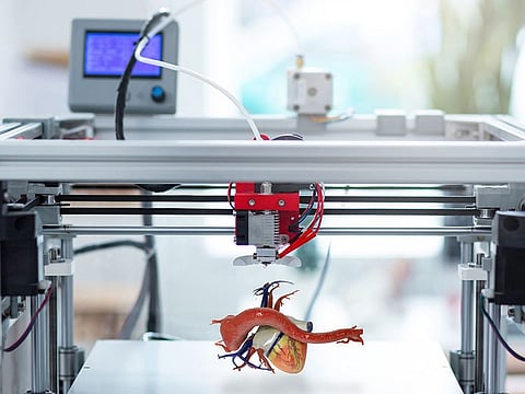
Stepping up to confirm the diagnosis and treat Shabbie, doctors at Cleveland Clinic decided to use a 3D printed model of a heart and its vessels.
Surgical intervention
The technology was developed by NYU Abu Dhabi’s technology platform, was subsequently employed to support surgical intervention and care for Shabbie.
As Dr. Yazan Aljabery, Cardiac Surgeon within the Heart, Vascular & Thoracic Institute at Cleveland Clinic Abu Dhabi, said, “Correcting a case of Kommerell’s Diverticulum when the aorta arches left, as is typical, is relatively straightforward because the deformity is accessible and visible. However, when the vessel arches right, as in this case, the defect is obscured by other large vessels, making surgical interventions particularly challenging. Using a 3D-printed model in such cases enhances the safety of the procedure and allows for more precise and tailored surgery.”
The process involved three stages; 3D image reconstruction, a procedure to create a three-dimensional model from raw diagnostic imaging data to provide a foundational digital framework of the patient’s anatomy; 3D slicing, which allowed for a more detailed analysis of the individual structures and organs in greater detail and; 3D printing, which involved building a physical replica of the patient’s anatomy that the surgeon could hold, examine and use for pre-surgical planning and simulation.
This technology is considered instrumental in understanding the unique challenges of each case and allows for a more precise and tailored surgical approach with the highest accuracy and minimum risks.

