Spot on!
Ask a surgeon about O-arm and all you'll get is praises for this technology. Find out how this piece of equipment, recently installed at Shaikh Khalifa Medical City in Abu Dhabi is redefining health care.
It is quite like crossing the final frontier, going where no man has gone before.
Imagine being able to look inside a living breathing human body and view it in all its wondrous detail. See, for instance, how a person's spine provides strong yet flexible support for the body even as it is protecting the delicate spinal cord within. Or view a person's knee cap from all angles to find out if it has sustained any injuries that might require surgery. And doing all this without having to move the patient from his bed!
While till recently all this belonged to the realm of sci-fi, today, computer assisted image-guided surgery (CAS) is offering surgeons the option of peering deep into the human body and viewing body parts from all possible angles to check on its condition and take any correctional steps if required.
And the piece of equipment which is making all this possible and making waves in the region among health professionals is the multidimensional imaging system called the O-arm.
Ushering a new era in advanced medical care, Shaikh Khalifa Medical City (SKMC) became the first clinical institution in the Gulf region to acquire this machine in May and several surgeries have been performed successfully utilising its calibre to optimum benefit.
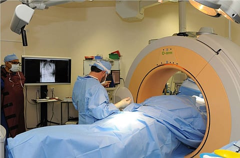
What it is
"The O-arm is a compact, robust, mobile lighter version of a CT-scanner,'' says Dr Peter Lange, senior consultant and Chair of Radiology at SKMC. "The ingenuity of the O-arm is that it is equipped with a [removable] gantry. It allows lateral access to the patient on the operating table and then closes in around him to offer 360-degree coverage. It is the only approved platform that provides surgeons with real-time, multi-plane standard fluoroscopy (an imaging technique used to obtain real-time moving images of a patient's internal structure using a fluoroscope) 2-D images and high-resolution 3-D images within the operating room.''
One of the main advantages of the O-arm is that "the potential for revision surgery is significantly reduced and complications are avoided simply because it permits accurate placement of hardware implants in the patient," says Dr Lange.
Orthopaedic spinal surgeon, Dr Veldsman has been applying his medical skill at SKMC for nearly six years now and has been one of the first surgeons in SKMC to utilise the O-arm to sterling effect. "The O-arm collects the data for us to be able to use the real stuff," he says. ‘The real stuff' for Dr Veldsman is hardware implants that he is required to ‘fit' into patients to enhance their quality of life. "Knee replacements, for instance, have been carried out for 50 years," Dr Veldsman points out, "but we can now position these implants much more accurately because we now have use of the O-arm.
"[That said] we use the O-arm mostly for spinal surgery,'' says Dr Veldsman.
"In certain spine-related conditions, we may have to insert implants into the spine,'' he says. The implants are held in place by screws and "we use a computer-assisted navigation system for accurate placement of these screws.'' It is here that the services of the O-arm come into focus.
The O-arm acquires a 3-D image of the spine which helps surgeons "insert screws at just the right places," Dr Veldsman continues. The image from the O-arm helps him view the spine from just about any angle, he says. "I can look at it from the top, the side, the bottom... That is real-time navigation," states Dr Veldsman with satisfaction.
The O-arm is linked to a navigation system. "For all practical purposes, the O-arm is out of the operating theatre.'' Once it takes the required images, it is moved out of the theatre. The images are loaded on to a computer which then links up with the navigation system. The doctors use this system to pinpoint locations in the body where corrections or surgery is required.
One of the main advantages of image-guided medical procedures is that such procedures are minimally-invasive as compared to open surgery for the same medical intervention.
Telling features of innovative O-arm
"Time is critical in the operating room," says Dr Lange.
The O-arm and the navigation system are two separate systems but are interlinked - the navigation system is based on images sourced by the O-arm.
The O-arm is better and more sophisticated than many of the imaging systems available today for several reasons, say experts. "For instance, after the surgery we can check the result using the O-arm while the patient is still on the operating table. We can check whether the hardware is placed properly. If it needs to be changed, that can be done immediately. We do not have to wait for a subsequent revision operation thus saving time in the long run," says Dr Lange.
Another advantage is that the patient need not be moved from the operating table for a new set of images to be taken. "We use the O-arm at the beginning of a [procedure] and at the end. If there is no space [in the operating theatre], we can remove the O-arm and [affix it later to the system] after the surgery. The O-arm is equipped with robotic re-positioning - in other words, it has a memory position store system so it knows exactly where to place itself in case we need images of the same location once again."
While previously the patient would have to be moved to the X-ray department after surgery if images were required, that is not necessary now thanks to the O-arm. "Thus with the installation of the O-arm in SKMC," says Dr Lange, "we can have immediate post-operative imaging done while the patient is still in the operating room.''
"Another possible post-operative situation is of a patient having to be moved to the Intensive Care Unit if he is in a critical condition and then being transported to the radiology department for imaging by the CT-scanner. That is now avoided."
The O-arm is incredibly time-effective. It takes just about 30 seconds to set it up. Says Dr Veldsman: "The actual radiation itself is done by the O-arm in 13 seconds. Not less than 391 images [giving a 360 degree view of the body part] are taken and saved in 13 seconds which can be called up at any time."
Less is more
Apart from knee replacement surgeries, SKMC surgeons will use the O-arm imaging system for operations that deal with spine and pelvic fractures, hip replacements, in cases of scoliosis, some complex cases of facial injuries as well as neurosurgery.
It will be of great use in 3-dimensional spinal deformities like scoliosis where the spine takes on an abnormal C or S curvature.
"The O-arm has a lot of value in cases that involve the cervical spine because there is much less space around it to manoeuvre for a surgeon," Dr Veldsman points out.
Ultimately, where spinal surgery is concerned, less - in terms of revision operations and complications - is more. For a spinal surgeon, every detail affects the accuracy of his work.


